
6 Best Microscope for Blood Analysis for Home Labs
Darkfield Blood Model Lab Darkfield & Brightfield Microscope is the top pick for most buyers. It earns that spot as the best microscope for blood analysis because its darkfield and brightfield modes support two common viewing approaches. That flexibility helps users compare clear, brightly lit slides with samples that benefit from darkfield illumination.
This guide is for students, hobbyists, educators, and clinicians choosing a microscope for prepared blood slides. The key factors include optical clarity, illumination control, objective magnification, and stage size. A stable frame also matters, since small movements can make slide work frustrating. The six picks below are ranked with clear reasons and honest drawbacks for each. You will find options for different budgets, skill levels, and workspace needs, from compact student models to larger laboratory-style instruments. Each review focuses on what the design can realistically support.
Quick Comparison
| # | Product | Best For | Price | |
|---|---|---|---|---|
| 1 | Darkfield Blood Model Lab Darkfield & Brightfield Microscope | Darkfield Blood Imaging Students | $599.40 | View → |
| 2 | Vabiooth Blood Version Compound Monocular Microscope | Entry-Level Blood Cell Observers | $297.40 | View → |
| 3 | AmScope T660C-DKO Trinocular Compound Microscope | Camera-Ready Lab Documentation | $951.48 | View → |
| 4 | Improved Hemocytometer Cell Counting Chamber, 2-Pack | Backup Cell Counting Workflows | $50.99 | View → |
| 5 | Improved Neubauer Hemocytometer Counting Chamber | Routine Manual Cell Counts | $11.99 | View → |
| 6 | JKSVTF X-S-Z-107D Monocular Biological Microscope | Basic School Biology Lessons | $405.30 | View → |
6 Top Microscopes for Blood Work
The list is ranked from sixth place down to the top pick, with each microscope’s features and limits explained.
6. JKSVTF X-S-Z-107D Monocular Biological Microscope

Best for: School lab slide work
The JKSVTF X-S-Z-107D is a biological microscope for viewing prepared animal, plant, and blood samples. It is listed at $405.30, placing it above many basic home microscopes. The product title calls it monocular, but the specifications describe a hinged binocular head tilted at 45 degrees. Buyers should confirm the supplied configuration before ordering.
Its four achromatic objectives provide 4X, 10X, 40X, and 100X magnification, with the 100X lens designed for oil immersion. A WF10X wide-field eyepiece is included, while a WF16X option is listed. The double-layer mechanical stage measures 140 by 140mm and moves 75mm by 45mm, giving users room to position slides accurately. Coaxial coarse and fine controls offer a 30mm focusing range. The fine adjustment scale reads 0.002mm, which supports careful focusing at higher magnification. These features fit structured classroom or home lab work better than casual viewing.
Pros
- Four achromatic objectives cover low, medium, and oil-immersion viewing.
- The 140 by 140mm movable stage supports controlled slide positioning.
- A 0.002mm fine-adjustment scale allows precise focus changes.
Cons
- The listing gives conflicting information about the head, calling the microscope monocular in the title and binocular in the specifications.
- Illumination details, included accessories, and the need for immersion oil are not clearly provided, so the $405.30 price is difficult to compare fairly.
5. Improved Neubauer Hemocytometer Counting Chamber

Best for: Routine microscope cell counting
This Improved Neubauer hemocytometer is a reusable glass counting slide for estimating cell density under a microscope. Its etched grid divides the viewing area into measured counting sections, allowing users to count cells or microorganisms in a known volume and calculate concentration. The design was originally made for blood-cell counts, but it can also suit other suspended cell samples when the sample and counting method fit the chamber.
At $11.99, its main appeal is a low-cost tool for repeated manual counts. High light transmission helps the sample remain visible through the glass, while clearly defined grid lines make chamber boundaries easier to locate. That matters when cells are small or numerous. The slide does not automate counting, so results still depend on even sample loading, correct focusing, and consistent counting rules. Buyers should confirm that their microscope can focus on the chamber and that they have the needed coverslip and pipette. As a reusable plate, it can reduce disposable chamber use, provided it is cleaned carefully after each session.
Pros
- Reusable glass construction supports repeated counting sessions.
- High light transmission helps keep cells visible.
- Defined grid lines support more consistent manual counts.
Cons
- Manual counting takes longer and can introduce user error.
- The listing does not provide chamber dimensions or grid specifications.
4. Improved Hemocytometer Cell Counting Chamber, 2-Pack

Best for: Manual cell counts under a microscope
This two-piece Neubauer hemocytometer set is designed for counting blood cells, other cell types, and microorganisms. Each chamber has two counting areas positioned in the center. Every large grid contains 16 medium grids, and each medium grid divides into 25 small grids. That creates 400 small grids per large grid, giving users a structured layout for manual counts. The chamber depth is 0.1 millimeters, while each small cell covers 0.0025 square millimeters.
The standout feature is the metal silver background with bright counting lines. Under a microscope, users can adjust image contrast to make those lines appear lighter or darker. That may help separate the grid from the sample during observation. The package includes four glass coverslips measuring 22 × 26 millimeters. This provides replacements for routine work, although users still need a compatible microscope, pipette, and suitable sample preparation method. The set does not automate counting or calculate results for you.
Pros
- Two counting areas support repeated counts or comparison between samples.
- The 400-small-grid layout provides a consistent counting pattern.
- Bright lines on a silver metal background allow contrast adjustment under a microscope.
Cons
- Counts remain manual, so results depend on careful loading, focusing, and calculation.
- The kit includes only four coverslips, which may run out during frequent laboratory use.
3. AmScope T660C-DKO Trinocular Compound Microscope
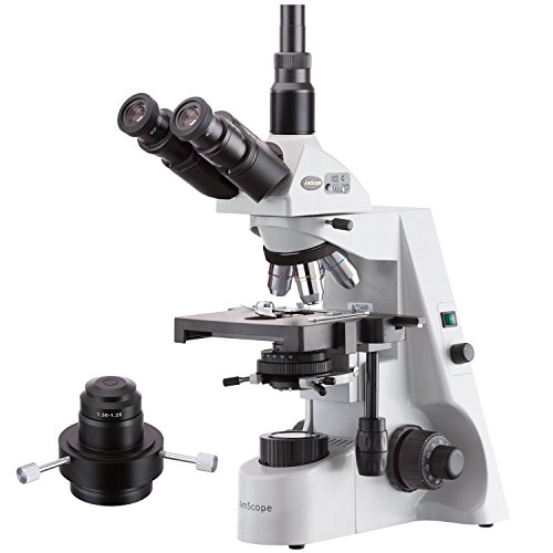
Best for: Advanced darkfield microscopy learners
The AmScope T660C-DKO is a professional compound microscope with a trinocular design and a 40x to 2500x magnification range. It offers six selectable magnification settings, giving users room to inspect prepared slides at low power and examine much finer details at higher power. The backward-facing nosepiece leaves more working space around the specimen, which can make slide handling less cramped during longer observation sessions. At $951.48, this is a serious investment for a home lab, classroom, or research-focused workspace.
Its lighting system is the main draw. Kohler halogen illumination works with a condenser and iris, allowing users to adjust how light passes through the sample. Brightfield lighting suits standard specimens, while darkfield illumination can make transparent or low-contrast objects easier to see against a darker background. The optical system is designed to produce sharp, well-defined views across the range. Still, 2500x magnification does not automatically reveal more useful detail if the slide or sample lacks enough resolution.
Pros
- Six settings from 40x to 2500x cover routine and high-power viewing.
- Brightfield and darkfield modes support different specimen types.
- The backward nosepiece provides more space around the slide.
Cons
- The $951.48 price may exceed many casual users’ budgets.
- Halogen lighting can produce more heat and use more power than LED systems.
2. Vabiooth Blood Version Compound Monocular Microscope

Best for: Live-blood hobbyists needing shared views
The Vabiooth Dual-View Compound Microscope is designed for viewing prepared slides and live-blood samples. It offers claimed magnification from 40X to 2500X through four achromatic objectives: 4X, 10X, 40X, and 100X spring-oil. A pair of wide-field glass eyepieces provides 10X and 25X viewing. The eyepieces sit at a 30-degree angle, which can make longer viewing sessions more comfortable.
Its main advantage is the second viewing path. A 5MP electronic eyepiece sends the image to a 7-inch LCD screen, giving several viewers a direct view without adding a separate monitor. The display rotates 360 degrees, which helps when sharing the image around a table. Its mechanical stage uses 1.0mm divisions and locks the slide in place while moving along the X and Y axes. That makes it easier to record and revisit a slide position. An aluminum carrying case protects the microscope and included accessories during storage or transport. Video and still capture are supported with a video capture card.
Pros
- The 7-inch LCD screen allows group viewing without an external monitor.
- The X/Y mechanical stage supports controlled slide movement and repeatable locations.
- Four objectives cover low-power scanning through oil-immersion viewing.
Cons
- The 100X objective requires immersion oil, adding cleanup and handling work.
- Video and photo capture require a separate video capture card, which adds cost beyond the $297.40 price.
1. Darkfield Blood Model Lab Darkfield & Brightfield Microscope

Best for: Live-blood microscopy and shared viewing
This compound microscope combines brightfield and darkfield viewing with an advertised 40X to 2500X magnification range. Its four achromatic objectives include 4X, 10X, 40X spring, and 100X spring-oil options. A 1.25 NA Abbe condenser with an iris diaphragm helps control illumination for different slides. The LED light provides a practical, low-maintenance source for routine observation.
The 5MP E-eyepiece feeds a built-in 7-inch LCD, so several people can view the sample without crowding around the eyepieces. The display rotates 360 degrees, and the 30-degree eyepiece angle can reduce neck strain during longer sessions. Three WF glass eyepiece options, 10X, 16X, and 25X, support different viewing setups. Its double-layer mechanical stage uses 1.0mm divisions and X-Y controls, allowing users to record slide positions and return to them later. At $599.40, the included aluminum case also adds useful protection for storage and transport.
Pros
- 5MP camera and 7-inch screen support group viewing.
- Mechanical stage offers 1.0mm position references.
- Four objectives cover low, medium, and oil-immersion magnification.
Cons
- Darkfield use is described specifically for live-blood research, not every specimen type.
- The 100X objective requires immersion oil and careful cleanup.
Buying Guide
Optical clarity is the most important factor in a microscope for examining blood. Clear lenses and even lighting reveal cell edges and details that cheap optics can blur or hide.
- Objective lenses: Choose a compound model with 4x, 10x, 40x, and 100x oil-immersion objectives. The 100x lens, paired with a 10x eyepiece, provides 1,000x magnification.
- Optical quality: Look for achromatic objectives, glass lenses, and a numerical aperture of at least 1.25 on the 100x objective. These features matter more than extreme advertised magnification.
- Illumination: Select a built-in LED with adjustable brightness and a color temperature near 5,000 to 6,500K. An Abbe condenser with an iris diaphragm helps control contrast.
- Mechanical stage: A metal stage with X-Y controls gives steadier slide movement than spring clips alone. Aim for at least 3 by 3 inches of travel.
- Accessories and cost: Budget $250 to $700 for a capable student or home laboratory setup. Confirm that it includes immersion oil, spare fuses, slide clips, and a 10-slide starter pack. Blood microscopy does not replace professional medical testing.
If you cannot get everything, prioritize the objective lenses and condenser before camera features or extra accessories.
Frequently Asked Questions
What type of microscope is best for examining blood cells?
A compound microscope with 400x to 1000x magnification is the most suitable choice for examining stained blood cells. Look for a mechanical stage, fine focus controls, and an adjustable LED light source. A 100x oil-immersion objective can reveal smaller details, but it requires prepared slides and immersion oil.
How much magnification do I need to view blood samples?
For routine blood-smear viewing, choose a microscope that reaches 1000x, with 400x useful for scanning the slide. Higher magnification alone does not add detail. Clear optics, steady focusing, and proper lighting matter just as much. Most users also need prepared slides and immersion oil for the 1000x objective.
Can a home microscope diagnose anemia or other blood conditions?
No, a home microscope cannot reliably diagnose anemia, infection, or other blood conditions. A clinician may need a properly prepared smear, blood counts, and other laboratory tests. Even a clear view at 1000x cannot identify every abnormality or explain its cause. Seek medical advice for symptoms or unusual results.
Are digital microscopes good for looking at blood samples?
Yes, digital microscopes can work well for viewing prepared blood smears, but image quality depends on optics, lighting, and camera resolution. Choose a model with at least 400x optical magnification and adjustable illumination. A camera can share images, but it does not turn the device into a diagnostic laboratory instrument.
Conclusion
Darkfield Blood Model Lab Darkfield & Brightfield Microscope is the top recommendation for buyers who want darkfield viewing and standard brightfield work in one setup. If your priority is routine brightfield viewing, choose a simpler brightfield microscope instead, since it may cost less and avoid features you may not use. Before buying, double-check the microscope’s overall size, compatibility with your slides and accessories, included quantity of lenses or prepared materials, and current price. Also confirm that the listing matches your intended use and provides clear setup information. A suitable optical system should fit your workspace and learning goals. Keep expectations realistic, since microscope images cannot replace clinical testing or professional interpretation.
